


Last week during a CBCT training on the Prexion Excelsior Pro, I came across a great example of the benefit of varying slice thickness in CBCT software. CBCT volumes show 3 different 2D perspectives (axial, sagittal, and coronal) of the same location. Because a CBCT volume is a composite of many "slices" in the same plane, CBCT x-ray allows the user to vary the thickness from a single voxel slice to a composite view of multiple slices in the same plane. A digital PA will look similar to a composite view of many slices.
The patient had pain in the number #19 region which had previously had a root canal. The first image is a PA of the region that was taken with a Scan-X PSP system. The second image is an axial CBCT image with the blue line representing the sagittal images that follow. The images following represent differing thicknesses along that blue line. Notice that the first thickness is .1mm thick and it has a high degree of graininess. This is true of any image that is a single voxel thickness. The last image is a composite of 128 slices and it looks similar to the PA with the lesion not being as apparent.
Getting to know how to vary slice thickness can give the clinician another helpful diagnostic view of the diagnosis at hand. Notice going from .1 mm slice to a 1.6mm slice shows the lesion is approximately the same diameter. This indicates that the lesion is approximately 2mm thick in the sagittal view. This, of course, could be verified with the axial view. Notice, also, that at 3.2mm thickness the distal buccal root comes into view.Typically, varying slice thickness is suggested to smooth the image and make it more readable. It is important to understand when to vary slice thickness and how doing so will create more of a composite x-ray.
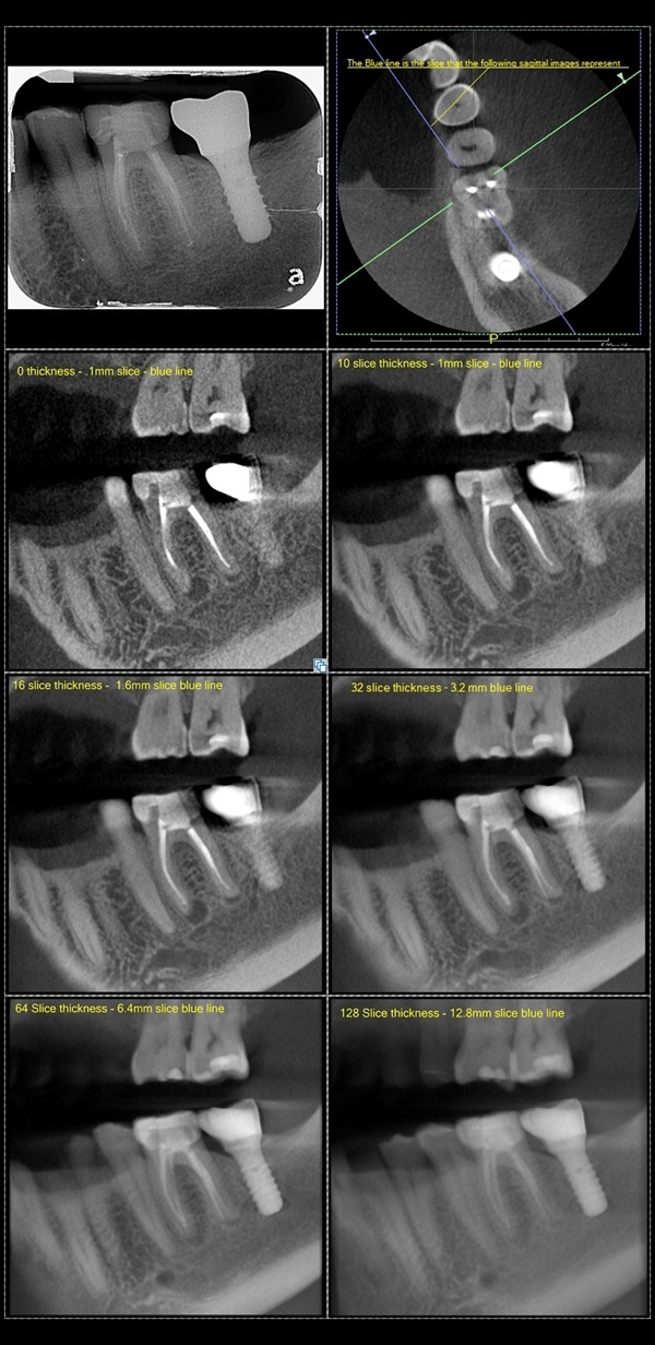
The following image helps explain slice thickness.
.jpg)
Leave Comment